Biophotons, cell metabolism and ultra-weak photon emission
The invisible light of life
Dr. Klaus Everding · Irina Outkine
We see light as something self-evident. Sunlight, a lamp, a screen – light is part of our everyday life. But even where it appears completely dark to our eyes, light can arise: in living cells.
This extremely weak light emission is referred to as ultra-weak photon emission (UPE). In colloquial terms, it is often called "biophotons." This refers to an emission of individual photons in connection with biochemical and metabolic processes. The amount of light is so small that it cannot be perceived by the human eye. However, modern highly sensitive cameras and photodetectors can measure it.
This opens up an unusual view of living systems: life is not only accompanied by chemical substances and electrical signals – weak electromagnetic processes can also leave measurable traces.
What are biophotons?
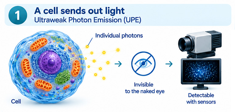
Fig. 1: Living cells can emit ultra-weak light. Own schematic representation.
Biophotons are not a mystical "light of life." In popular science usage, the term primarily describes the ultra-weak light emission of biological systems. According to the current state of research, its origin is primarily associated with oxidative and metabolic processes.
Reactive oxygen species, or ROS for short, play an important role. They are produced normally in cell metabolism, including in the mitochondria. In certain reaction chains, molecules can reach excited states. When these states are exited, a photon can be emitted.
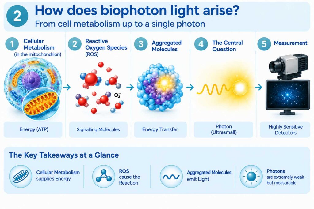
Fig. 2: Simplified pathway of UPE formation: Metabolic processes can lead to photon emission via reactive oxygen species and excited molecules. Own representation after Mould et al. (2024).
ROS are by no means fundamentally harmful. In normal concentrations, they belong to regular signaling and defense processes. Only when production and degradation become unbalanced can oxidative stress occur.
The UPE is interesting because it may be related to changes in cell metabolism and physiological state. Whether it plays a functional role itself beyond that is a much more open question.
How weak is this light?
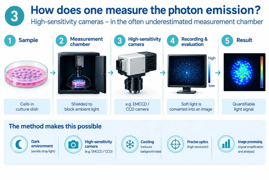
Fig. 3: Principle of UPE measurement using highly sensitive CCD/EMCCD technology. Own schematic representation.
The intensity of ultra-weak photon emission lies far outside our normal sensory perception. A study published in 2025 cites approximately 10 to 10³ photons per square centimeter per second in the range of about 200 to 1000 nanometers for biological systems.
For comparison: An ordinary lamp emits so much visible light that our eyes can work with it without any problems. In contrast, UPE involves individual, extremely rare photons. Even the smallest disturbances – such as ambient light, thermal radiation, or electronic signals – can influence the measurement.
This explains why the investigation is technically demanding: one needs heavily darkened measurement environments, sensitive detectors, and careful data evaluation.
From the Gurwitsch effect to Fritz-Albert Popp
The idea that living organisms could emit a form of light or react to light is not new. An early important name is the Russian biologist Alexander Gurwitsch, who described evidence of radiation emanating from living tissue as early as the 1920s.
From the 1970s onwards, the German biophysicist Fritz-Albert Popp in particular dealt intensively with ultra-weak photon emission. Popp went beyond pure measurement. He hypothesized that under certain conditions, biophotons might not only be by-products of metabolism but could also be involved in information transfer and regulation.
This is where the area begins where a clear scientific separation is particularly important.
The existence of ultra-weak photon emission is well documented. In contrast, the more far-reaching ideas about the special coherence of biophotons and biological information transfer are not established in the same way. Questions regarding reproducibility, physical coherence, and specific biological mechanisms remain the subject of scientific discussion.
Popp is therefore interesting primarily because he asked questions that are experimentally verifiable – not because all of his hypotheses are considered proven.
What modern cameras make visible
The technology used already shows how difficult the measurement is. Highly sensitive CCD and especially EMCCD cameras can capture individual or very few photons.
A study published in 2025 by Salari and colleagues, for example, compared living and dead mice. The living animals showed a significantly higher photon emission. In plants, too, the UPE changed under stress such as injury or increased temperature.
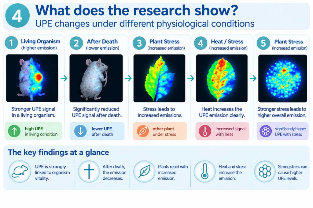
Fig. 4: Simplified representation of the findings reported by Salari et al. (2025): Differences between living and dead mice as well as changes in UPE during plant stress. Own representation; no reproduction of the original figure.
The result must not be overinterpreted. It does not mean that plants or animals emit "pain light" or that a mysterious life energy becomes visible here. Rather, it shows that physiological states and stresses can be associated with measurable changes in ultra-weak photon emission.
This makes UPE a possible research tool: perhaps something about the state of a biological system can be read from the light emission.
Biophotons as biomarkers?
If certain changes in cell metabolism are reliably reflected in the UPE, photon emission could serve at least as an indirect marker for certain biological processes.
Initially, that would not be spectacular at all. Science often works with measurable accompanying phenomena to investigate processes that are themselves difficult to access. The decisive factor would therefore not be whether biophotons are "healing" or "informative," but whether their intensity, their temporal course, or their spectral distribution are reproducibly related to certain biological states.
Stronger statements require controlled experiments, independent replications, and standardized measurement procedures.
METAVITAL and the classification of MNLS technology
A separate line of development is pursued by METAVITAL. According to its own statements, the company has been developing and selling products and systems based on biophoton or MNLS technology for diagnostic and therapeutic applications for more than a decade.
For a professional classification, it is explicitly stated: The MNLS method and the corresponding systems are to be assigned to complementary or alternative medicine. Their methods and effects have not yet been recognized as scientifically proven in conventional medicine or natural science.
METAVITAL also points this fact out itself.
At the same time, METAVITAL reports numerous documented successes from application. Such experience reports are not scientifically equivalent to controlled clinical studies. However, like interesting experimental findings, they can be a reason for further research.
It is precisely at this point that the connection to the biophoton studies described here becomes interesting. If evidence of biological effects of light and electromagnetic processes continues to consolidate through independent, reproducible research, the scientific basis for applications such as MNLS technology could also broaden.
What was scientifically investigated at METAVITAL?
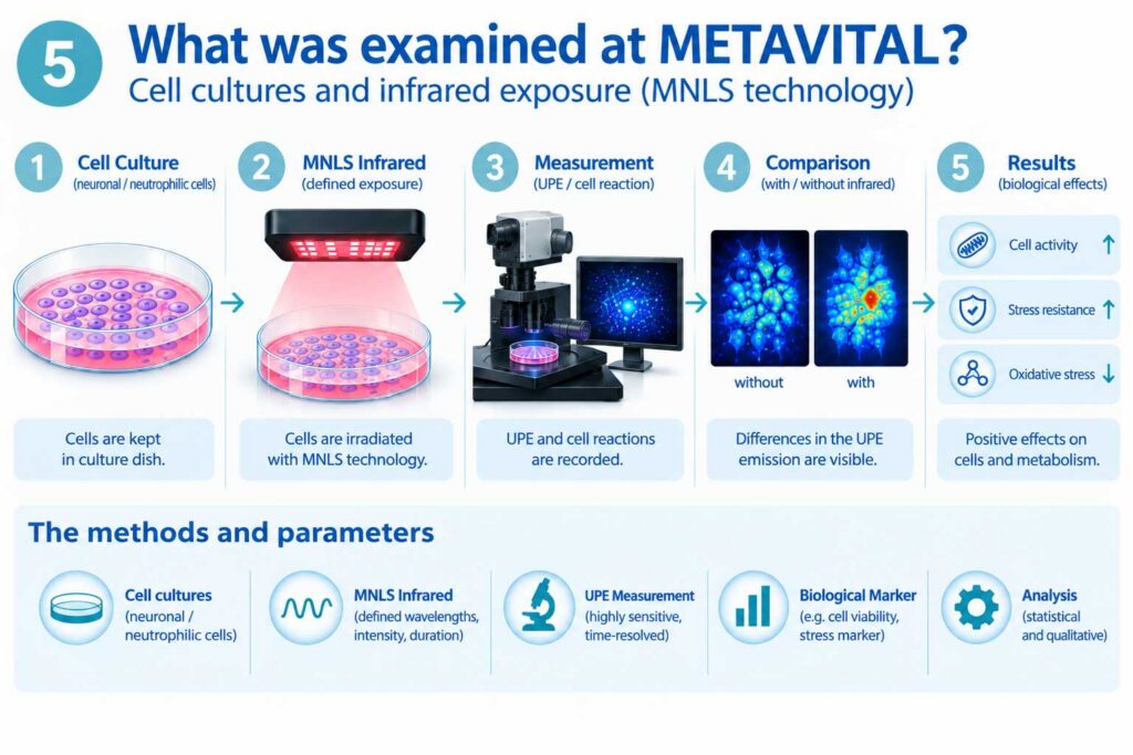
Fig. 5: Schematic representation of the in vitro experiment with cell cultures and infrared application. The study measured cell parameters, not UPE directly. Own representation.
In January 2026, the Dartsch Scientific Institute investigated human cell cultures in vitro on behalf of METAVITAL. Among others, SH-SY5Y cells, a neuronal cell model, and differentiated HL-60 cells with neutrophil-like properties were examined.
A sensor with a light intensity sensor and an infrared source was used. Irradiation took place under controlled conditions; a single exposure duration of 60 minutes was derived from preliminary tests.
In the SH-SY5Y cells, a 10.1 ± 3.3 percent higher vitality compared to the untreated control was reported in the XTT test after the single 60-minute application. In addition, stronger regeneration or cell migration was described.
In the differentiated HL-60 cells, the formation of endogenous radicals was investigated. Here, an 18.8 ± 4.4 percent stronger reduction in radical formation was shown compared to the control group; the stated statistical value was p ≤ 0.01.
These results are interesting as experimental observations. However, they prove neither therapeutic efficacy in humans nor a connection with biophotons. In the study, ultra-weak photon emission was not measured directly.
This is crucial: A change in cell parameters after an infrared application is not automatically proof that biophotons mediate the effect.
The open key question
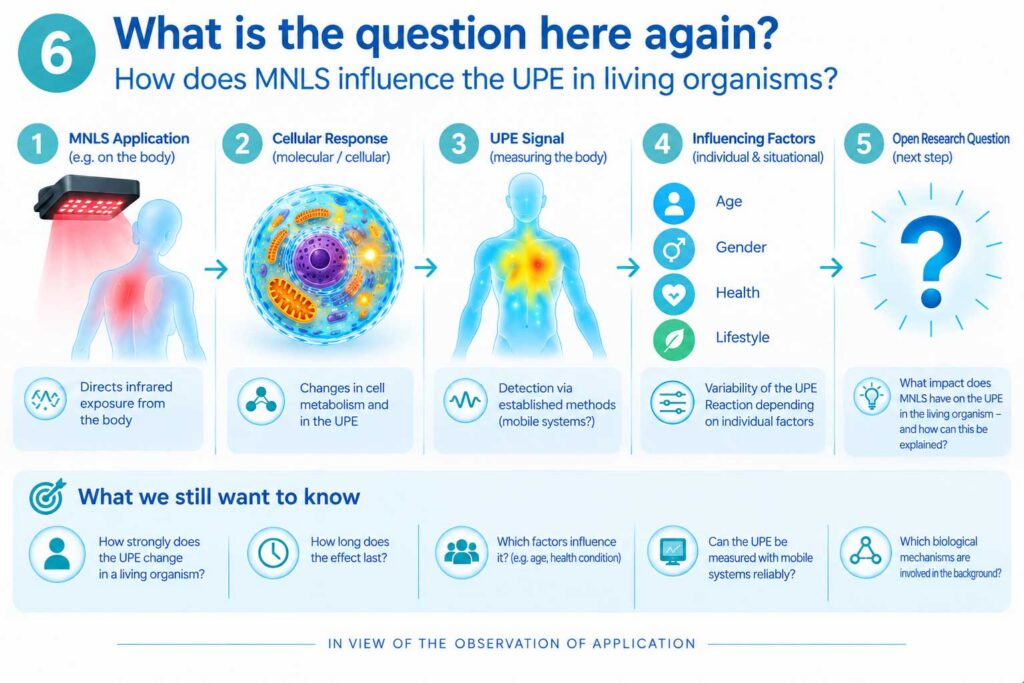
Fig. 6: The central open research question: Does an MNLS application change the ultra-weak photon emission of living cells? Own schematic representation.
This is precisely why a next experimental step would be particularly exciting.
Cell cultures could be divided into a treated and an untreated or sham-treated group under strictly controlled conditions. Subsequently, it could be investigated with calibrated photodetection or an EMCCD camera whether the ultra-weak photon emission changes as a result of the MNLS application.
The intensity of the emission, its temporal course, its spatial
distribution and – as far as technically possible – its spectral composition would be of interest.
This would allow a specific question to be asked:
Does an MNLS application change the ultra-weak photon emission of living cells?
If so, the next step would be the question of the mechanism. Is the effect due to the infrared radiation itself, to a specific modulation, to thermal or photochemical processes – or to an interplay of several factors?
These would no longer be philosophical questions. They could be measured.
Light as a new window on life?
The various research directions meet at a common horizon without claiming the same mechanism.
The UPE research shows that living systems emit extremely weak light and that this emission can be related to metabolism and physiological changes. The work in the environment of Fritz-Albert Popp has also raised the question of whether light in biological systems could possibly be more than a passive by-product.
The investigations into MNLS technology, in turn, follow a different direction: here, infrared is applied to cells from the outside to investigate possible biological reactions.
Whether there is a direct connection between these phenomena is currently open.
This is exactly why the question is scientifically interesting. Science does not begin where everything is already explained, but where an observation can be transformed into a verifiable question.
What is already certain today
Some points are relatively clear:
1
Living biological systems emit ultra-weak light.
2
The emission is closely related to oxidative and metabolic processes.
3
Modern detectors can capture this extremely weak radiation.
4
Changes in the physiological state can be accompanied by changes in the UPE.
5
Whether UPE also has an independent function in communication or regulation is open.
6
The cell culture effects investigated on behalf of METAVITAL are interesting in vitro findings, but not proof of clinical efficacy and not proof of a biophoton effect.
Perhaps this is the most exciting perspective: The “light of life” does not have to be mysterious to be scientifically fascinating.
A single photon is initially just a photon. But when controlled measurements show when it arises, how its emission changes, and whether biological systems react to it, a tiny light signal can become a new window on the physics of life.
And perhaps the next interesting investigation begins exactly where a simple question stands: If a technology acts on living cells – does it also change their own ultra-weak light?
Then one would no longer have to speak of “light of life.”
It could be measured.
Sources and further reading
Mould, R. R. et al. (2024): Ultra weak photon emission—a brief review. Frontiers in Physiology.
Salari, V. et al. (2025): Imaging Ultraweak Photon Emission from Living and Dead Mice and from Plants under Stress. Journal of Physical Chemistry Letters 16, 4354–4362.
Prasad, A.; Pospíšil, P. (2013): Towards the two-dimensional imaging of spontaneous ultraweak photon emission from microbial, plant and animal cells. Scientific Reports 3, 1211.
Dartsch Scientific (2026): In vitro study on the effect of infrared application on cultured human cells, on behalf of METAVITAL.
METAVITAL: Information on MNLS technology, biophoton technology and
classification as a complementary/alternative medicine method.
Image rights and source notice:
This article was created with the participation of Chat GPT. The six figures in this version are own schematic representations. The scientific statements in Fig. 2 and Fig. 4 are based on the respective sources mentioned. The underlying review by Mould et al. (2024) is published Open Access under CC BY; a direct reproduction of their figure was deliberately not used here.